In cancer diagnostics, the old question was what a tumour is. Increasingly, the harder question is where its cells are, whom they are talking to, and whether the surrounding tissue is helping the disease resist treatment. MultivisionDx, a Helsinki-based University of Helsinki spin-out founded in April 2025, has built its case on that shift: cancer prognosis, it argues, can be read not only from malignant cells but from the spatial ecology of the tumour and its microenvironment.
The company describes its mission as “turning biological complexity into clinical clarity”, positioning itself at the intersection of AI image analysis, spatial biology and clinical decision support for solid cancers. Its technology uses computer vision to integrate tumour-cell features, stromal composition and tissue architecture into patient-specific tumour profiles intended to flag patients at risk of failure under standard-of-care treatment.
At the centre of the company’s scientific pitch is a 2024 Cell paper, “Multiparameter imaging reveals clinically relevant cancer cell-stroma interaction dynamics in head and neck cancer”, led by Sara Wickström’s research group and co-authored by MultivisionDx founders Karolina Punovuori and Fabien Bertillot. The study reported an image-analysis pipeline that combines cell-state markers with morphology and spatial context, identifying clinically relevant tumour phenotypes in head and neck squamous cell carcinoma, or HNSCC.
The unmet need is substantial. The Cell paper notes that HNSCC remains aggressive and difficult to treat, with five-year overall survival around 50 per cent, recurrence in about 50 per cent of patients during the first two years after diagnosis, and median overall survival below one year after failure of first-line therapy. The authors also argue that standard clinicopathological variables, including tumour site, nodal involvement, tumour thickness and margin status, only weakly predict recurrence, survival and treatment response.
MultivisionDx’s core proposition is that tissue images contain more clinically useful information than current diagnostic workflows extract. On its technology page, the company says multiplex immunofluorescence can capture 20 to 50 protein markers in a single tissue scan while preserving spatial context across the tumour microenvironment, after which its AI-based workflow performs cell phenotyping, spatial network mapping and biomarker scoring.
The scientific novelty is not simply the use of AI on pathology images. Many digital pathology tools aim to automate tasks pathologists already perform, such as counting positive cells or standardising histological grading. MultivisionDx’s claim is more ambitious: that its platform can produce new biological information by combining protein expression, cellular morphology, neighbourhood relations and stromal architecture at single-cell resolution.
In the Cell study, researchers first analysed pathologist-curated tumour microarrays from 212 HNSCC patients with clinical data, using multiplexed immunofluorescence panels to quantify epithelial markers, differentiation states, stemness markers and partial epithelial-mesenchymal transition, or pEMT, markers. The pipeline segmented nuclei and cytoplasms, measured marker intensity, nuclear shape and local neighbourhood features, then clustered cells to generate phenotypic signatures at patient level.
The first striking result was that epithelial tumour phenotypes alone told only part of the clinical story. The researchers identified six epithelial signatures, including Sox2-high groups, well-differentiated tumours and pEMT-related patterns. These signatures correlated with histopathological features, HPV-positive oropharyngeal cancers, metastasis and recurrence patterns, but they did not strongly predict overall or disease-specific survival in primary tumour biopsies.
The stronger signal emerged when the team looked at the stroma, the non-malignant tissue environment surrounding and infiltrating tumours. The study identified three broad stromal signatures: immune-enriched stroma, CAF-enriched fibrotic stroma, and myeloid-high stroma. Patients with immune-enriched stroma had five-year disease-specific survival of 64 per cent, compared with 34 per cent for those with CAF-enriched fibrotic stroma and 52 per cent for those with myeloid-enriched stroma.
Yet the most provocative finding was combinatorial. In tumours with a pEMT epithelial signature, stromal identity dramatically altered outcome: five-year disease-specific survival reached 94 per cent for patients with immune-enriched stroma, but only 21 per cent for those with CAF-enriched stroma. In non-pEMT tumours, stromal composition was far less decisive.
That result is the basis for MultivisionDx’s “compound biomarker” concept. Rather than treating tumour-intrinsic markers and microenvironmental features as separate diagnostic layers, the company seeks to integrate cell phenotypes, tissue architecture and neighbourhood interactions into a single prognostic score. Its diagnostic product page describes an in-development IVD-grade spatial biomarker test for treatment selection in HNSCC and other solid cancers.
The mechanistic story matters because it makes the biomarker more than a black-box classifier. In the Cell paper, spatial transcriptomics on eight independent tongue squamous cell carcinoma samples suggested that pEMT cancer cells and CAF-enriched stroma formed a hub of intercompartmental signalling, particularly through extracellular matrix pathways and AREG-EGFR interactions. Follow-up co-spheroid experiments with patient-derived cancer cells and cancer-associated fibroblasts showed that CAFs could reprogramme pEMT-like cancer cells towards a pro-invasive phenotype.
The authors went further, showing that the invasive behaviour depended on cancer-cell state and EGFR signalling. UTSCC74 cells, which had stronger EMT and EGFR-pathway features, responded to CAF co-culture with transcriptional reprogramming and invasion, while UTSCC76 cells did not show the same response. In 3D collagen assays, CAFs substantially enhanced invasion of UTSCC74 cells, and gefitinib, an EGFR inhibitor, blocked invasion of both cancer cells and CAFs in the co-culture model.
For a clinician or policy audience, the key point is that MultivisionDx is not merely proposing a more detailed microscope. It is trying to convert spatial cell biology into a risk stratification product: identify patients whose tumours appear biologically more aggressive than conventional staging suggests, and guide escalation, de-escalation or alternative therapy decisions accordingly. The company’s diagnostic product page says its lead test is intended for HNSCC, where current staging fails to identify patients who may benefit from treatment de-escalation or intensification.
The commercial path is still early. MultivisionDx says its platform originated in a Business Finland Research to Business project, received €680,000 in Business Finland funding to prepare commercialisation, and became a company in April 2025. It later raised a €1 million pre-seed round in December 2025 led by Antler, with participation from Helsinki University Funds, Kaikarhenni Oy and a Finnish angel investor in healthcare.
The company has also undergone a leadership transition. Karolina Punovuori, the founding CEO and lead author of the Cell paper, moved into the role of Chief Scientific Officer after Michael Wittinger was appointed CEO in November 2025. Wittinger brought diagnostics, oncology and regulatory-sector experience, including previous work at Platomics, where the company says he helped scale the organisation from 20 people to more than 100.
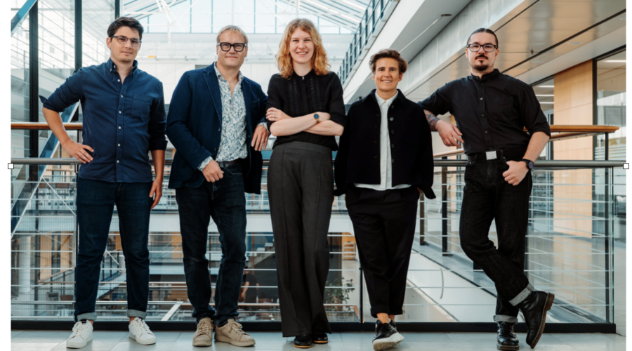
The founding team reflects the hybrid nature of the project. MultivisionDx lists Michael Wittinger as CEO, Karolina Punovuori as Chief Scientific Officer, Fabien Bertillot as Head of Computational Biology, János Lengyel as Head of Engineering and Sara Wickström as Science Advisor. Wickström is Director of the Max Planck Institute for Molecular Biomedicine and a research director and principal investigator at the University of Helsinki.
The company’s near-term regulatory roadmap is explicit but ambitious. Its diagnostic product is currently described as research use only, with the platform validated in the 650-patient study, followed by planned in-house clinical use under Article 5.5 in 2027 and full EU IVDR Class C certification from 2028 onwards.
That timeline underlines the central caveat. The Cell paper itself states that the patient cohorts were retrospective biobank samples and that future prospective studies are needed to validate the predictive nature of the compound biomarkers. It also notes that its Visium spatial transcriptomics lacked single-cell resolution, meaning annotated spots included mixtures of cell types and could not support more granular transcriptional profiling of every population.
This is where the policy question becomes sharper. If validated prospectively, tools like MultivisionDx could help health systems allocate intensive treatment where it is most likely to help, avoid toxic overtreatment where biology suggests lower risk, and identify patients for trials or targeted therapies. But if adopted prematurely, spatial AI diagnostics could add cost, complexity and algorithmic opacity to oncology pathways without clear evidence of improved outcomes.
The company appears aware of that translational gap. It says it is working with international clinical centres to validate its diagnostic test in multi-site studies before market entry, while University of Helsinki material says the company aims to integrate its technology into major digital pathology platforms as an add-on module and produce kits for staining tumour biopsies.
There is also a platform ambition beyond HNSCC. Health Incubator Helsinki reported that MultivisionDx is already applying the technology to a 2,000-patient colorectal cancer cohort, while the company says its architecture is designed for expansion into additional solid tumours where spatial biology adds predictive value beyond current standard of care.
The strategic attraction for hospitals, investors and pharmaceutical partners is clear. For hospitals, a validated spatial biomarker could refine treatment selection. For drug developers, it could help identify patient subgroups whose tumours are driven by particular microenvironmental interactions. For investors, it sits in a fast-growing convergence zone between AI, pathology, oncology and precision diagnostics.
The risk is equally clear: translating a compelling retrospective biological discovery into a regulated, reproducible, reimbursable clinical product is difficult. Multiplex staining must be standardised, image pipelines must survive real-world variation across scanners and laboratories, and clinicians must be able to interpret the resulting reports with enough confidence to alter treatment plans. MultivisionDx’s planned clinician-friendly prognostic report and IVDR pathway are designed to address precisely those hurdles, but validation will decide whether the promise holds.
For now, MultivisionDx is one of the more interesting examples of a broader movement in precision oncology: away from single biomarkers and towards ecological diagnostics. Its thesis is that cancer is not only a mutation-bearing clone but a living tissue system, and that the arrangement of malignant cells, immune cells, fibroblasts and extracellular matrix can reveal who is likely to relapse, resist treatment or benefit from a different course.
The most telling line comes from Sara Wickström, quoted after the Cell study won Finland’s Medix Prize: “certain combinations of malignant cells and tissue cell types in supposedly healthy surrounding tissue have a strong prognostic effect on the progression of cancer”. That is the story MultivisionDx must now prove at clinical scale: that the neighbourhood of a tumour can become a diagnostic readout, and that reading it well can change a patient’s path.
References
- Health Incubator Helsinki. (2025, August 12). MultivisionDx’s diagnostic tests match cancer patients with therapies that actually work. Health Incubator Helsinki. [healthincu…lsinki.com]
- Helsinki Innovation Services, University of Helsinki. (2025). MultivisionDx offers a patient-specific ‘fingerprint’ for cancer diagnostics. University of Helsinki. [helsinki.fi]
- MultivisionDx. (2026). MULTIVISIONdx. [multivision.ai]
- MultivisionDx. (2026). Technology. [multivision.ai]
- MultivisionDx. (2026). Diagnostic product: Spatial diagnostic test. [multivision.ai]
- MultivisionDx. (2026). Team. [multivision.ai]
- MultivisionDx. (2025, October 6). MultivisionDx scientists win prestigious Finnish medical research prize. [multivision.ai]
- MultivisionDx. (2025, November 14). MultivisionDx appoints Michael Wittinger as new CEO. [multivision.ai]
- MultivisionDx. (2025, December 3). MultivisionDx secures €1M pre-seed funding round to transform cancer diagnostics. [multivision.ai]
- Punovuori, K., Bertillot, F., Miroshnikova, Y. A., Binner, M. I., Myllymäki, S.-M., Follain, G., Kruse, K., Routila, J., Huusko, T., Pellinen, T., Hagström, J., Kedei, N., Ventelä, S., Mäkitie, A., Ivaska, J., & Wickström, S. A. (2024). Multiparameter imaging reveals clinically relevant cancer cell-stroma interaction dynamics in head and neck cancer. Cell, 187(24), 7267–7284. https://doi.org/10.1016/j.cell.2024.09.046 [Multiparam…eck cancer | PDF]
- Tech Funding News. (2025, December 2). Exclusive: MultivisionDx nabs €1M to decode cancer ‘fingerprints’ with AI.