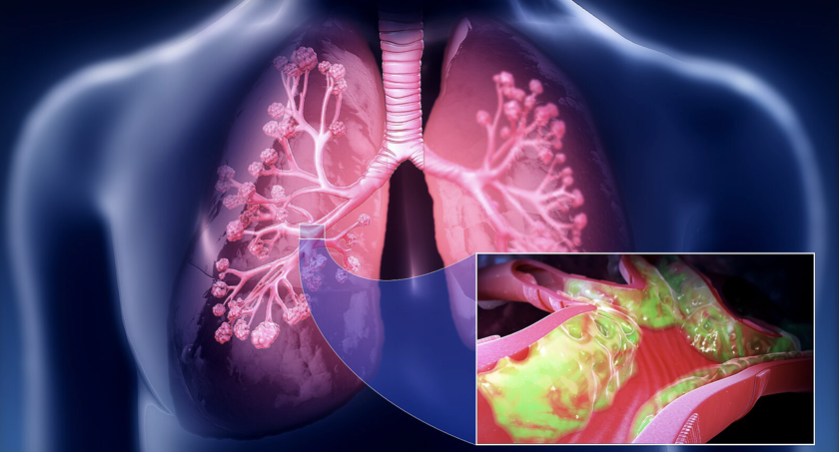
Kristel Svalland Knudsen’s PhD thesis, The lower airways microbiome in patients with interstitial lung diseases(University of Bergen, 2025), synthesizes a decade of work across two rigorously executed studies (MicroCOPD and MicroILD). Using protected bronchoscopy sampling and next‑generation sequencing (16S rRNA, ITS for fungi, and whole‑genome shotgun metagenomics), the thesis draws a coherent picture: the lower airways in interstitial lung diseases (ILDs), notably idiopathic pulmonary fibrosis (IPF) and sarcoidosis, harbour distinct microbial communities with reduced diversity and altered antimicrobial peptide (AMP) profiles. These signatures could inform diagnosis, stratification, and, ultimately, tailored interventions.
For clinicians, researchers, and policy makers, the work is notable for its methodological stringency (protected sampling, negative controls, decontamination pipelines) and for bridging microbiome profiles with host immunity (AMPs). For the general public, the takeaway is straightforward: the lungs are not sterile; their resident microbes might influence how certain lung diseases begin, progress, and respond to treatment.
Key Findings
1) IPF vs. COPD and healthy controls
- Microbial diversity: IPF shows lower alpha diversity in protected BAL (PBAL) and protected brushings versus COPD and controls.
- Taxonomy: IPF lower airways dominated by Firmicutes and Bacteroidetes (with Actinobacteria third), whereas COPD shows relatively more Proteobacteria.
- Microaspiration signal: Greater similarity between oral wash and lower airway communities in IPF suggests an oropharyngeal contribution (microaspiration/reflux).
- Host defense: Lower hBD‑1 (and trending lower SLPI) in IPF PBAL; in IPF, hBD‑1 inversely correlates with Firmicutes (driven by Streptococcus), hinting at disease‑specific host–microbiome interactions.
2) Sarcoidosis vs. controls
- Fungal signal: Enrichment of Aspergillus in PBAL among sarcoidosis patients; bacterial differences are subtle.
- Diversity: Bacterial alpha diversity is lower in sarcoidosis (OW and PBAL); both bacterial and fungal beta diversity differ from controls.
- Host defence: Lower SLPI and hBD‑1 in PBAL, compatible with impaired mucosal defence and/or an inactive/treated disease phenotype.
3) Unclassifiable ILD (metagenomics)
- Community profiles: Distinct beta‑diversity compared with healthy controls; partial overlap with IPF or sarcoidosis.
- Biomarker concept: A microbial dysbiosis index (DI) derived from species‑level metagenomics discriminates ILD groups from controls and helps refine the profile of unclassifiable ILD, an area of high unmet need.
Why This Matters Clinically and Strategically
Diagnostic precision and stratification
- Reduced diversity and characteristic taxa (plus DI) could support MDD (multidisciplinary discussion) in separating IPF, sarcoidosis, and unclassifiable ILDs—especially when imaging is equivocal or tissue is risky to obtain.
Therapeutic angles
- AMP deficits (e.g., SLPI, hBD‑1) point toward host‑directed therapies, adjunctive AMP‑based treatments, or strategies to modulate airway microbiota (antimicrobials, probiotics, targeted anti‑fungals).
- The microaspiration link in IPF strengthens the case for aggressive reflux assessment and management, though antifibrotic benefits from anti‑reflux therapy remain debated; microbiome endpoints could offer new ways to measure impact beyond FVC.
Public health and policy
- Standardized protected sampling and bioinformatic decontamination are resource‑intensive but crucial. Policies that fund and mandate methodological consistency (sampling, primers, databases, analysis pipelines) will improve reproducibility and accelerate translation.
Strengths, Gaps, and Next Steps
Strengths
- Protected sampling (PBAL/PSB) with negative controls, gold‑standard in low biomass sites, reduces upper airway carryover.
- Parallel assessment of host AMPs and microbiota makes the work mechanistically richer than taxonomy‑only surveys.
- Inclusion of healthy controls and cross‑disease comparisons (COPD, sarcoidosis, IPF) adds context.
Limitations
- Cross‑sectional design limits causal inference—are microbial changes drivers or consequences?
- Low biomass and host DNA contamination complicate shotgun metagenomics; species assignments (e.g., Staphylococcus) can reflect cross‑mapping or lab contaminants if not exhaustively controlled.
- Sample size constraints reduce power for differential abundance and confounder adjustment; disease activity and treatment status (especially in sarcoidosis) can blur signals.
- Taxonomy > function: 16S/ITS infer identity, not functional capacity (e.g., biofilm formation, virulence, metabolite pathways). Function would sharpen therapeutic hypotheses.
What should happen next
- Longitudinal cohorts with repeated protected sampling, integrating clinical endpoints (exacerbations, lung function decline, survival) to test prognostic value of diversity/taxa/DI.
- Functional profiling (metagenomics/metatranscriptomics/metaproteomics) to connect taxa with pathways (e.g., fibrogenic signaling, AMP response, biofilm).
- Interventional trials, anti‑reflux strategies, targeted anti‑fungals (where Aspergillus signatures persist), AMP analogues, and microbiome modulation—powered with microbiome endpoints.
- Standardization. shared protocols, mock communities, multi‑lab proficiency testing, curated reference databases, to enable meta‑analyses and policy guidance.
Takeaways
- For clinicians: consider reflux rigorously in IPF; be open to microbiome‑informed risk stratification when the evidence base matures.
- For researchers: pair protected sampling with functional omics; treat contamination and compositionality as first‑order design constraints.
- For policy makers: fund standardization and longitudinal infrastructure; prioritize biobanking and interoperable bioinformatics.
- For the public: lung health includes the microbiome; lifestyle and co‑morbid conditions (e.g., reflux) may influence microbial ecology and disease course.
References
Bolyen, E., Rideout, J. R., Dillon, M. R., et al. (2019). Reproducible, interactive, scalable and extensible microbiome data science using QIIME 2. Nature Biotechnology, 37(8), 852–857.
Clarke, E. L., et al. (2018). Microbial lineages in sarcoidosis: A metagenomic analysis tailored for low‐microbial content samples. American Journal of Respiratory and Critical Care Medicine, 197(2), 225–234.
Dickson, R. P., et al. (2017). Bacterial topography of the healthy human lower respiratory tract. mBio, 8(1), e02287-16.
Hiemstra, P. S., Amatngalim, G. D., van der Does, A. M., & Taube, C. (2016). Antimicrobial peptides and innate lung defenses: Role in infectious and noninfectious lung diseases and therapeutic applications. Chest, 149(2), 545–551.
Knudsen, K. S., et al. (2022). The lower airways microbiome and antimicrobial peptides in idiopathic pulmonary fibrosis differ from chronic obstructive pulmonary disease. PLOS ONE, 17(1), e0262082.
Knudsen, K. S., et al. (2022). The lower airways microbiota and antimicrobial peptides indicate dysbiosis in sarcoidosis. Microbiome, 10, 175.
Knight, R., et al. (2018). Best practices for analysing microbiomes. Nature Reviews Microbiology, 16(7), 410–422.
Molyneaux, P. L., et al. (2014). The role of bacteria in the pathogenesis and progression of idiopathic pulmonary fibrosis. American Journal of Respiratory and Critical Care Medicine, 190(8), 906–913.
Raghu, G., et al. (2018). Diagnosis of idiopathic pulmonary fibrosis: An Official ATS/ERS/JRS/ALAT clinical practice guideline. American Journal of Respiratory and Critical Care Medicine, 198(5), e44–e68.
Tipton, L., Ghedin, E., & Morris, A. (2017). The lung mycobiome in the next-generation sequencing era. Virulence, 8(3), 334–341.
Xia, G. H., et al. (2019). Stroke Dysbiosis Index (SDI) in gut microbiome is associated with brain injury and prognosis of stroke. Frontiers in Neurology, 10, 397. (Applied analogously for dysbiosis index concept.)